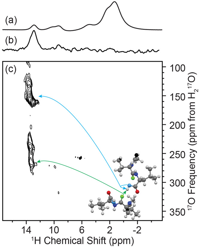
Figure 7.
MAS NMR spectroscopy of N-Ac-VL: (a) 1H direct Hahn-echo detection, (b) one-dimensional 1H–17O R3-R-INEPT, (c) and two-dimensional 1H–17O R3-R-INEPT spectrum with R3 = 100 μs. Spectra were acquired with ωR/2π = 20 kHz at 17.6 T (ω0H/2π = 750 MHz). Correlations between the leucine carbonyl proton to its directly bonded oxygen (blue arrow) and its next closest oxygen through space (NCO, green arrow) are indicated on the crystal structure of N-Ac-VL in the inset.92

