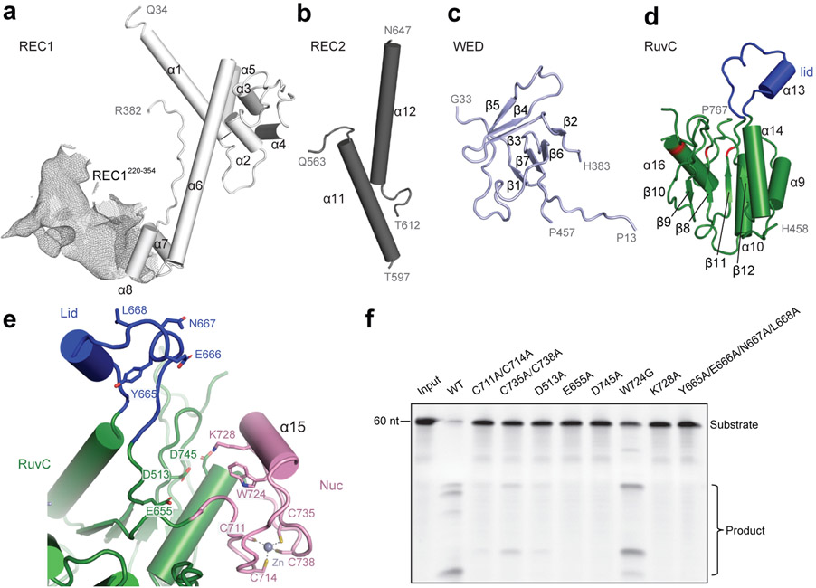
Figure 2. Domain structure and active site of Cas12g.
a-d, Cartoon presentation of the REC1 domain(a), REC2 domain(b), WED domain(c), and RuvC domain(d). Density of REC1220-354 observed by 3D focused classification is shown in mesh. The triplet of acidic residues in the active site and the lid motif colored in red and blue, respectively. e, The endonuclease center in the RuvC domain. The acidic residues D513, E655, and D745 are labeled. The Nuc domain is attached to the active center, with K728 and W724 pointing to the active site. A zinc ion chelated by four cysteines (C711, C714, C735 and C738) is present in the Nuc domain. f, Substrate RNA cleavage assay using wild-type Cas12g and Cas12g with mutations as indicated. The results shown are representative of three experiments.

