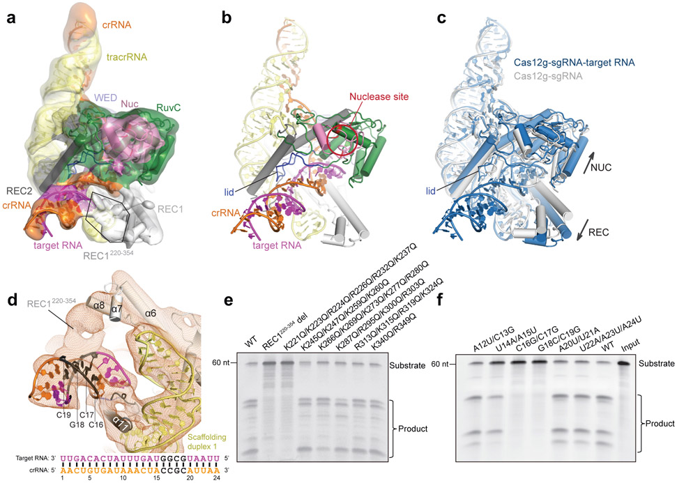
Figure 5. Target RNA recognition of Cas12g.
a, Cryo-EM map and model of the Cas12g-sgRNA-target RNA complex with each domain of Cas12g color-coded as in Fig. 1a. The crRNA, tracrRNA, and target RNA are colored in orange, light yellow, and magenta, respectively. b, Cartoon presentation of the Cas12g-sgRNA-target RNA complex. c, Structural superimposition of Cas12g-sgRNA (grey) and Cas12g-sgRNA-target RNA (sky blue) complexes. Conformational changes in Cas12g domains and sgRNA after recognition of target RNA are indicated by arrows. d, Contact between REC1220-354 and positions 16-19 of the crRNA-target RNA duplex. Schematic of the crRNA spacer-derived guide and target RNA is shown below. e, Substrate RNA cleavage assay using wild-type Cas12g and Cas12g with REC1220-354 deleted or with group mutations within REC1220-354. f, Substrate RNA cleavage assay using wild-type and mutant target RNAs. The results in e and f are representatives of three experiments.

