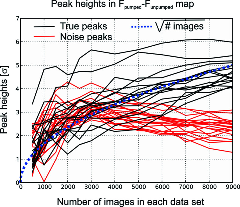
Figure 3.

The development of peak heights in an |F pumped| − |F unpumped| difference map derived from SFX data as a function of the number of images. The data used are from bacteriorhodopsin, 33 ms after light excitation (Nass Kovacs et al., 2019 ▸), to a resolution of 1.8 Å. Using the maps calculated with 3000 and 9000 images in both datasets, 15 ‘real’ peaks caused by structural change on light excitation were chosen, as well as 18 ‘false’ (spurious) peaks that could not be distinguished from the true signal when 3000 images were used for each dataset. Both positive and negative peaks were selected, and the negative peaks inverted so as to have positive values. The heights of these peaks (black: true peaks, red: false peaks) were plotted as a function of the number of images used for both datasets. The dashed blue line is proportional to the square root of the number of images.
