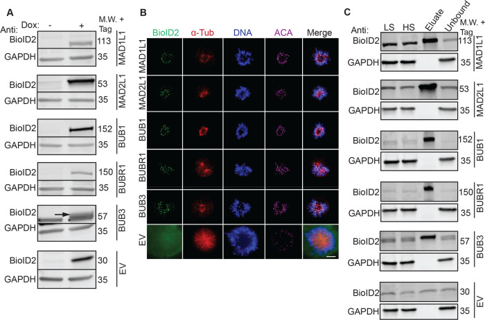
Figure 2.
Establishment of inducible BioID2-tagged SAC protein (BUB1, BUB3, BUBR1, MAD1L1, and MAD2L1) stable cell lines and biochemical purifications. (A) Immunoblot analysis of extracts from doxycycline (Dox)-inducible BioID2-tag alone (EV, empty vector) or BioID2-tagged SAC protein (BUB1, BUB3, BUBR1, MAD1L1, MAD2L1) expression cell lines in the absence (−) or presence (+) of Dox for 16 h. For each cell line, blots were probed with anti-BioID2 (to visualize the indicated BioID2-tagged SAC protein) and anti-GAPDH as a loading control. M.W. indicates molecular weight. Note that BioID2-tagged SAC proteins are only expressed in the presence of Dox. The arrow points to the induced BioID2-BUB3 protein band and the asterisk denotes a nonspecific band recognized by the anti-BioID2 antibody. (B) Fixed-cell immunofluorescence microscopy of the BioID2-tag alone (EV) or the indicated BioID2-tagged SAC proteins during prometaphase, a time when the SAC is active. HeLa BioID2-tagged protein expression cell lines were induced with Dox for 16 h, fixed and stained with Hoechst 33342 DNA dye and anti-BioID2, anti-α-Tubulin, and anticentromere antibodies (ACA). Bar indicates 5 μm. Note that all BioID2-tagged SAC proteins localize to the kinetochore region (overlapping with the ACA signal), whereas the BioID2-tag alone (EV) was absent from kinetochores. (C) Immunoblot analysis of BioID2 biochemical purifications from cells expressing the indicated BioID2-tagged SAC proteins or the BioID2-tag alone (EV). For each cell line, blots were probed with anti-BioID2 (to visualize the indicated BioID2-tagged SAC protein) and anti-GAPDH as a loading control. M.W. indicates molecular weight, LS indicates low-speed supernatant, and HS indicates high-speed supernatant. Uncropped immunoblots are provided in Figures S17 and S18.

