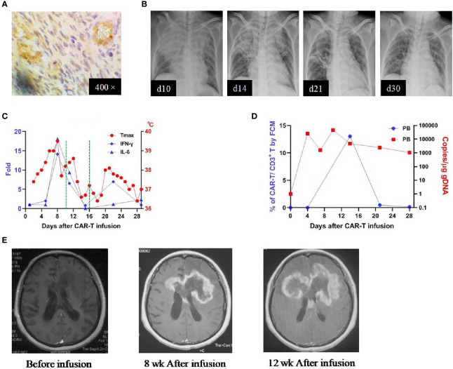
Figure 2.
IHC staining of the tumor and clinical response in patient 1. (A) Anti-EphA2 IHC staining of tumor tissue before enrollment. (B) Thoracic X-ray performed on days 10, 14, 21, after the infusion of CAR T-cells, showing the development and resolution of pulmonary edema in the right superior and middle lobes. (C) Dynamic changes of critical cytokines in the peripheral blood along with temperature, after the infusion of CAR T-cells. The dotted green line on the x-axis indicating the time window for the use of dexamethasone. (D) Expansion of CAR T-cells in the peripheral blood by flow cytometry (left y-axis) and qPCR (right y-axis). (E) Contrast-enhanced MRI performed before and after (8 and 12 weeks) the infusion of CAR T-cells.

