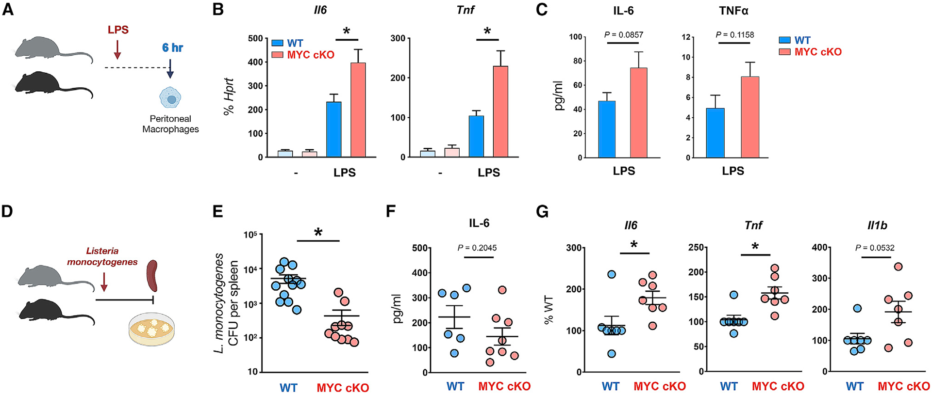
Figure 4. Myeloid-specific MYC-deficient mice exhibit augmented inflammatory responses and ameliorated clearance of bacteria in vivo.

(A) A schematic diagram illustrating the experimental design in (B) and (C). Peritonitis was induced in 6-week-old WT or MYC cKO mice by intraperitoneal injection of LPS (100 ng/mice).
(B) The mRNA expression of Il6 and Tnf in peritoneal macrophages at 6 h after LPS injection (Control, n ≥ 3; LPS injection, n ≥ 6).
(C) Concentrations of IL-6 and TNF-α in the serum at 6–9 h post-infection (n = 12). p = 0.0857 (IL-6) or 0.1158 (TNF-α) by two-tailed, unpaired t test.
(D) A schematic diagram illustrating the experimental design in (E)–(G). 12-week-old WT and MYC cKO mice were intravenously infected with Listeria monocytogenes.
(E) Bacterial titers in the spleen were determined at day 3 post-infection (n ≥ 10).
(F) Concentrations of IL-6 in the serum at 22–24 h post-infection (WT, n = 6; MYC cKO, n = 8). p = 0.2045 by two-tailed, unpaired t test.
(G) The mRNA expression of Il16, Tnf, and Il1b (% of WT) in peritoneal macrophages at 22 h post-infection (n = 7).
All data are shown as mean ± SEM. *p < 0.05 by one-way ANOVA with a post hoc Tukey test (B) or two-tailed, paired t test (E and G).
