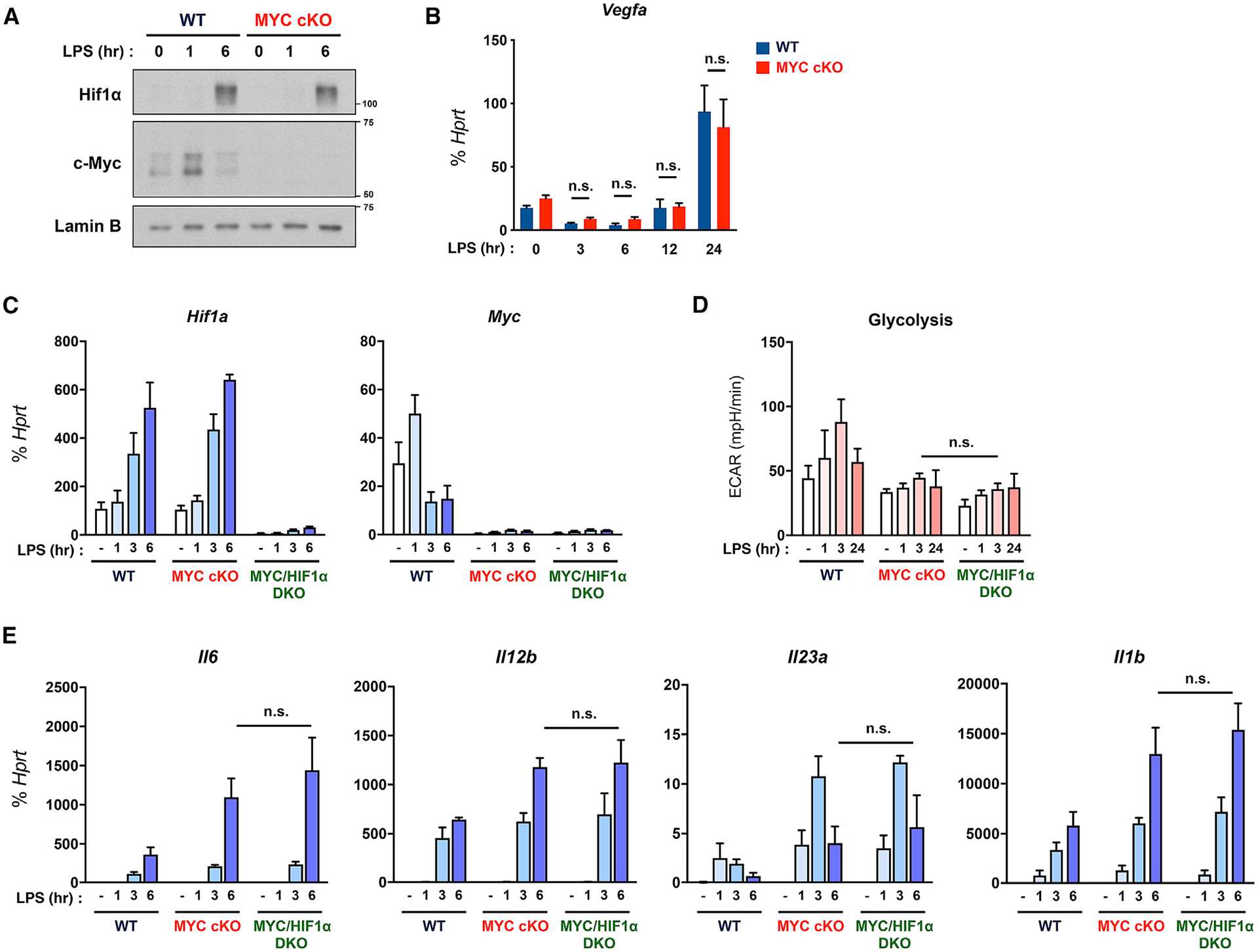
Figure 6. MYC regulates early LPS responses independent from HIF1α.

(A) The expression of MYC and HIF1α was determined by immunoblot by using nuclear lysates in WT and MYC cKO BMDMs after LPS (10 ng/ml) stimulation for the indicated time points. Lamin B served as a loading control. Data are representative of 4 experiments.
(B) The mRNA expression of Vegfa in WT and MYC cKO BMDMs after LPS (10 ng/ml) stimulation for the indicated time points (n = 3).
(C) The mRNA expression of Myc and Hif1a in WT, MYC cKO, and MYC/HIF1α-double deficient (DKO) BMDMs after LPS (10 ng/ml) stimulation for the indicated time points (n = 3).
(D) The quantified glycolysis from seahorse glycolysis stress tests in WT, MYC cKO, and MYC/HIF1α DKO BMDMs after LPS (50 ng/ml) stimulation for the indicated time points (n ≥ 3).
(E) The mRNA expression of Il6, Il12b, Il23a, and Il1b in WT, MYC cKO, and MYC/HIF1α DKO BMDMs after LPS (10 ng/ml) stimulation for the indicated time points (n ≥ 4).
All data are shown as mean ± SEM. n.s., not significant by two-way ANOVA with a post hoc Tukey test.
