
Fig. 1. Cellular source of IL-33 controls the outcome of parasite helminth infection.
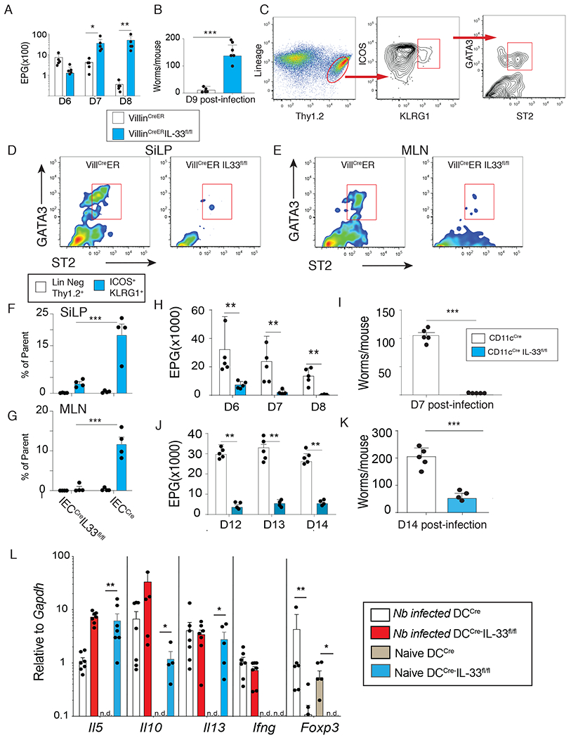
(A-B) VillinCreER or VillinCreER IL-33fl/fl mice were given tamoxifen (i.p. 1 mg/mouse daily starting day −1) and subcutaneously infected with 750 L3 Nippostrogylus brasiliensis (N.b.). Fecal egg counts on day 6-8 (A) and adult worm counts on d9 (B) post-infection. (C) Gating strategy to identify Lin−Thy1.2+GATA3+ ST2+ innate lymphoid cells (ILC2) (d9 post-infection). Lineage markers: B220, CD3, CD5, CD11b, CD11c, CD19, and NK1.1. (D-E) Representative flow plots showing ST2+ ILC2 in and small intestine lamina propria (SiLP) (D) mesenteric lymph nodes (MLN) (E) in VillCreER vs VillCreERIL-33fl/fl. (F) Quantification of Lin−Thy1.2+GATA3+ ST2+ from the Lin−Thy1.2+ gate (open bars) or the Lin−Thy1.2+ICOS+KLRG1+ gate (blue bars) from VillinCreER and VillinCreERIL-33fl/fl strain in (F) SiLP and (G) MLN. (H-I) Fecal egg counts on d6-8 (H) and adult worm counts on d7 (I) after N.b. infection in CD11cCre (open bars) vs CD11cCreIL33fl/fl mice (blue bars). (J) Fecal egg counts on d12-14 (K) and adult worm counts on d14 after Heligmosomoides polygyrus bakeri (H.p.b.) infection in CD11cCre vs CD11cCreIL33fl/fl mice. (L) Levels of Il5, Il10, Il13, Ifng, and Foxp3 in jejunum of CD11cCre vs CD11cCreIL33fl/fl at the steady state of d9 post-N.b. infection assessed by quantitative real-time PCR. Representative results from 3 independent experiments. Data show mean ± SEM with each symbol represents one mouse. * p< 0.05, ** p< 0.01, and *** p< 0.001 as determined by Student’s t-test.