
Fig. 4. DC express the pore-forming protein Perforin-2 at the plasma membrane in an IL-33/ST2-dependent manner.
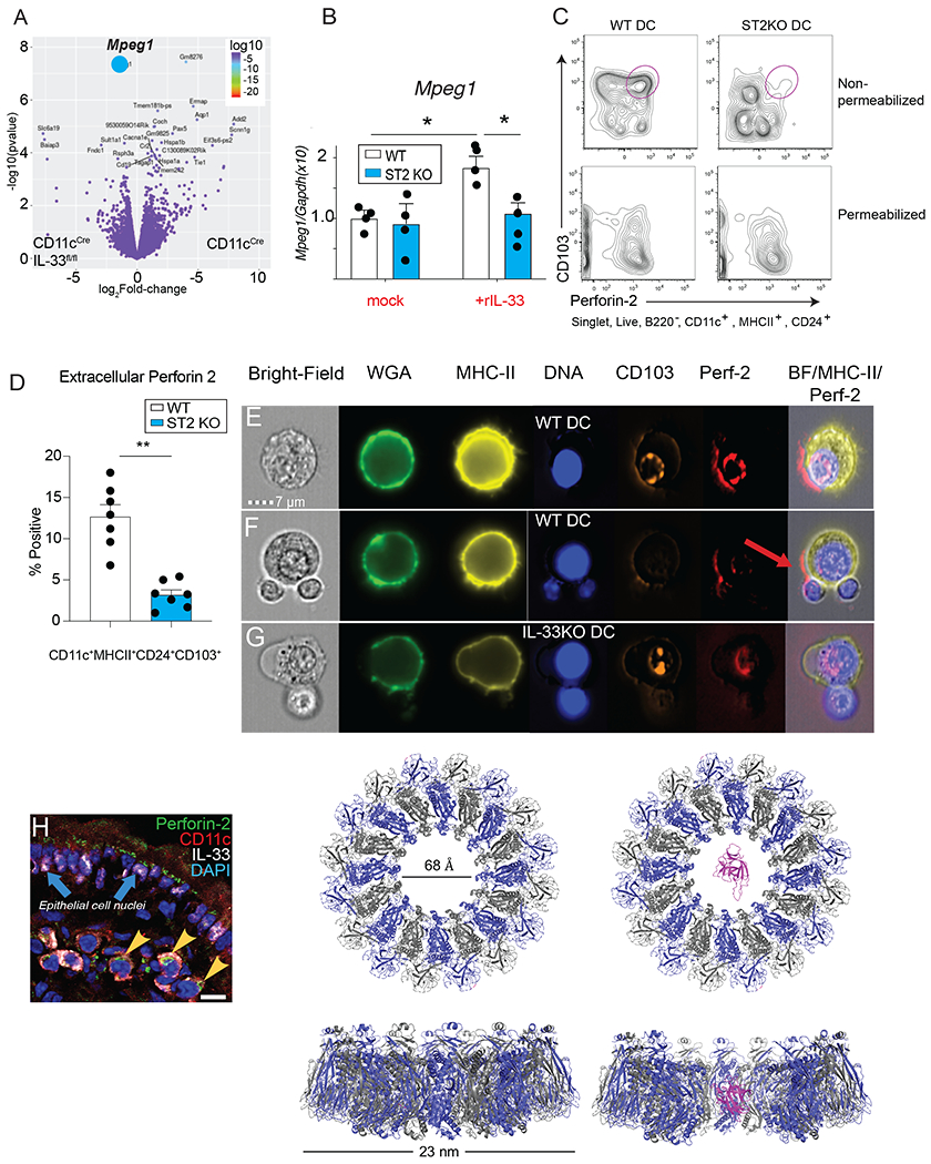
(A) Volcano plot showing differential gene expression in flow-sorted CD103+ DC from MLN of CD11cCre vs CD11cCreIL-33fl/fl mice. The blue symbol indicates Mpeg1. (B) Transcript levels of Mpeg1 in BMDC from WT vs ST2−/− treated with or without recombinant IL-33 for 24h, * p< 0.05 as determined by ANOVA. (C) Representative flow plots showing Perforin-2 staining on WT vs ST2−/− BMDC with or without permeabilization. (D) Quantification of intact Perforin-2 positive CD103+ BMDC from WT vs ST2−/− mice in experiment described in “B”. Representative results from 2 experiments n=3-4/group. (E-F) OT-II cells were co-cultured with WT or (M) IL-33−/− BMDC under Treg-differentiating conditions (DC:T= 1:4) for 3d, surface stained with specific reagents to visualize single channel and merged images for brightfield, wheat germ agglutinin, HLA-DR, CD103, Perforin-2 and analyzed with Amnis ImageStream® flow cytometry at 60X magnification. Red arrow indicates the distribution of Perforin-2 on the surface of DC. (H) Immunofluorescent staining of Perforin-2 (green), CD11c (red), and IL-33 (white) on mouse small intestine. Blue arrows indicate IL-33 expression in epithelial cells and yellow arrowheads indicate co-staining of IL-33, CD11c, and Perforin-2, (scale bar= 15 microns). (I) Model of human interleukin-33 (magenta) placed in the pore of wild-type Perforin-2. Perforin-2 is a hexadecamer with alternate subunits colored grey and blue. Model generated with Pymol using PDB ID 2KLL for IL-33 and PDB ID 6U23 for MPEG-1/perforin-2.