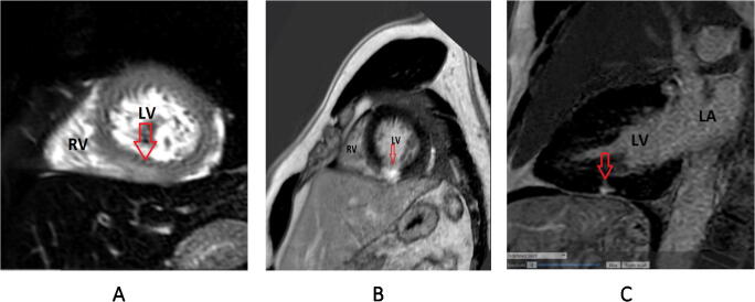
Fig. 2.
A Cardiac MRI: T2-weighted images showed myocardial edema of the inferior wall, suggestive of an acute myocardial injury (red arrow points to myocardial edema in image A) (from Am J Med. 2020 Aug; 133(8):e425-e426) [42]. B and C Post contrast images showed a focal area of inferior subendocardial late gadolinium enhancement (red arrows point to scar in images B and C). (From Am J Med. 2020 Aug; 133(8):e425-e426) [42]

