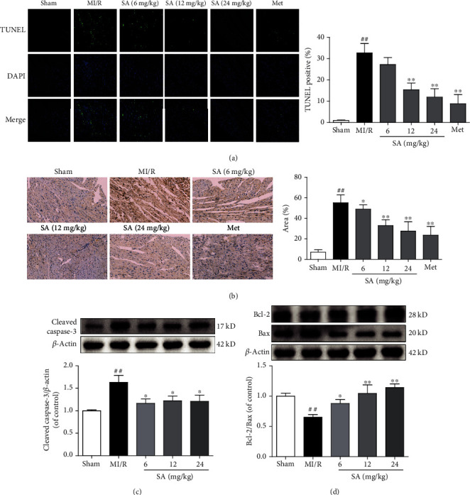
Figure 3.

SA inhibited cardiomyocyte apoptosis in MI/R mice. (a) Representative images of TUNEL staining (bar = 10 μm). (b) Immunohistochemical staining of cleaved caspase-3 (bar = 100 μm). (c) The expression of cleaved caspase-3 was detected by western blot. (d) The expressions of Bcl-2 and Bax were detected by western blot. The data were expressed as the mean ± SD. ##P < 0.01 vs. sham group; ∗P < 0.05 and ∗∗P < 0.01 vs. MI/R group (n = 3).
