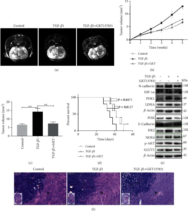
Figure 7.

Inhibition of NOX4 suppressed tumorigenesis in xenograft mice. (a) The representative image of intracranial tumors of mice was monitored and measured by MRI. (b) Tumor volume monitored by MRI. (c) The tumor volume was measured by MRI. (d) The percentage of the number of mice remaining after they received intracranial injections of either control or TGF-β1 alone or GKT137831 in combination at the indicated doses. (e) Western blot for the indicated proteins of intracranial tumors from the mice. (f) HE staining of control, TGF-β, and TGF-β/GKT137831 groups, and the tumor borders as well as infiltrating and invasion of the tumor are shown (N: normal brain tissue; T: tumors in brain; scale bar = 100 μm). Data represent mean and SD of three independent experiments. ∗P < 0.05; ∗∗P < 0.01; ∗∗∗P < 0.001.
