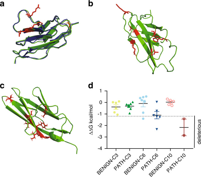
Fig. 4. Structural analysis of pathogenic missense MYBPC3 variants.

MyBP-C (the protein encoded by MYBPC3) domains C3, C6, and C10 were structurally modeled using I-TASSER33–35 (PyMOL,cartoon, green). Wild-type residues that are affected by missense pathogenic variants are depicted in red (PyMOL, sticks). (a) For the C3 domain, the I-TASSER model is aligned with an available NMR structure (2mq0.pdb,28 blue, PyMOL cartoon). Pathogenic variants within C3 largely cluster in a surface-exposed region. (b) C6 domain and (c) C10 domain pathogenic variants do not cluster within a specific region of the domain. (d) Results of STRUM19 analysis for MYBPC3 pathogenic and benign variants within C3, C6, and C10 are shown, with mean and SEM for each group depicted. Graph is labeled to indicate variants predicted to be deleterious.
