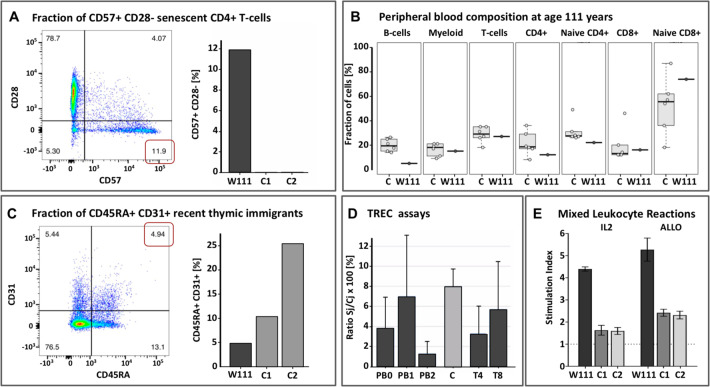
Fig. 2. Immune characterization of W111.
A Left: Flow cytometry Sorting of peripheral blood taken at age 110 showed increased fractions of CD57+ CD28- senescent CD4+ and CD8+ T-cells relative to middle aged controls, as apparent by the expression of CD57; Right: Proportion of senescent cells in W111 compared to middle-aged controls C1 and C2. B Proportions of sorted immune subsets (B-cells, Myeloid, T-cells, CD4+ T-cells, naive CD4+ T-cells, CD8+ T-cells, naive CD8+ T-cells) in peripheral blood of six middle-aged female controls (left) in W111 at age 111 years (right). C. Left: At age 110 years, nearly 5% of the CD4+ T-cells expressed both CD45RA and CD31, indicative of recent thymic emigrants; Right: The level of recent thymic emigrants in W111 was compared to middle-aged controls C1 and C2 [D] The percentage of T-cell receptor excision circles (TRECs) in in W111’s peripheral blood and sorted CD4+ and CD8+ T-cells at age 110 and 111 years was comparable to that of middle-aged healthy female controls C (3–6%). PB: Peripheral Blood cells; T4: CD4+ T-cells; T8: CD8+ T-cells. Numbers signify time points: 0: age 103; 1: age 110; 2: age 111. E In vitro proliferation assays: we computed Stimulation Indices for an IL2/TCR-dependent (IL2) and an allogeneic mixed-lymphocyte assay (ALLO) assay of cultured T-cells of W111 and two middle-aged female controls C1 and C2. In both assays, T-cells collected from W111 outperformed those taken from middle-aged controls on a per cell basis. Furthermore, see Supplement for flow cytometry analyses of cells collected at age 110 and 111, which indicated that both the CD4+ and CD8+ T-cell subsets contained considerable fractions of in vivo activated cells, evidenced by their high CD25 expression and CD69 expression.

