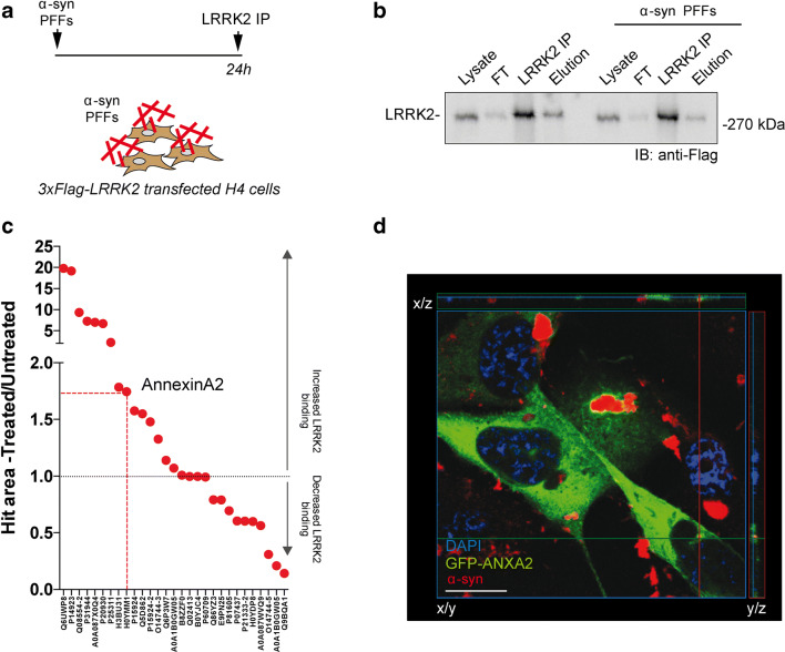
Fig. 5.
Characterization of LRRK2 interactoma in stimulated condition. A Schematic outline of the experimental setup. H4 cells were transfected using 3xFlag-LRRK2 encoding plasmid and, after 48 h post transfection, treated with 0.5 μM sonicated unlabeled α-syn PFFs for 24 h. LRRK2 was subsequently immunopurified using anti-Flag agarose beads (IP), eluted with Flag peptide (elution), and subjected to LC-MS/MS analysis. FT, flow-through; unbound LRRK2. B Western blot analysis showing LRRK2 expression, immunopurification, and elution in H4 cells in treated and basal conditions. C Relative quantification of LRRK2 interactome under treated and untreated conditions. The area of the precursor ions identified by LC-MS/MS analysis was used as a quantitative measure of the protein content. The ratio between the area of the precursor ions of untreated and treated samples (normalized by the content of LRRK2) was then considered to highlight proteins showing a different affinity for LRRK2 in the two conditions (n = 2). D H4 cells transfected with GFP-ANXA2 in α-syn PFF-treated condition verifying the proximity of internalized α-syn fibrils and transfected AnxA2. AnxA2-GFP (green), α-syn (red), DAPI (blue). Scale bar 30 μm

