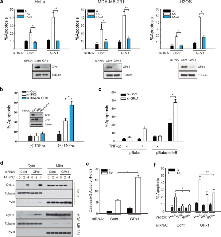
Fig. 1. GPx1 suppresses the TNF-α-induced apoptosis of RIPK3-negative cancer cells.
a HeLa, MDA-MB-231, and U2OS cells were transfected with a mixture of three siRNAs specific to GPx1. After 6 h of treatment with cycloheximide (C) alone or TNF-α plus cycloheximide (T/C), the cells were labeled with propidium iodide (PI) and annexin-V and subjected to FACS analysis. The pan-caspase inhibitor z-VAD-fmk (Z) was added as a pretreatment for 1 h before T/C stimulation. The data in the graph are the means ± SD of the percentage of apoptotic cells (n = 3, **P < 0.005, **P < 0.001, and #P < 0.0001). Firefly luciferase-specific siRNA was used as a control (Cont). b, c IKKβ-depleted (b) or super-repressor IκBα (srIκB)-expressing HeLa cells (c) were transfected with siRNA (#3) against GPx1 and treated with TNF-α (10 ng/ml) for 8 h. The data in the graph are the means ± of the percent of apoptotic cells (n = 3, *P < 0.001). The knockdown levels of the GPx1 and IKKβ proteins were verified by immunoblotting. d HeLa and MDA-MB-231 cells were transfected with GPx1 siRNA (#3) for 36 h and subjected to subcellular fractionation following T/C stimulation. Cytosolic (Cyto) and mitochondrial (Mito) fractions were subjected to immunoblot analysis for determining cytochrome c (Cyt.c) release. A representative blot is shown (n = 3). Tubulin and peroxiredoxin III (PrxIII) are cytosolic and mitochondrial markers, respectively. e siRNA-transfected HeLa cells were stimulated with T/C and lysed for use in a caspase-3 activity assay. The data in the graph are the means ± SD of the fold change of caspase-3 activity (n = 3, *P < 0.0001). f Vector control (Puro)-, Bcl-2-, and Bcl-xL-expressing HeLa cell lines were transfected with either control or GPx1 siRNA, stimulated with T/C for 8 h, and subjected to apoptosis assays. The data in the graph are the means ± SD of the percent of apoptotic cells (n = 3, *P < 0.001 and **P < 0.0001).

