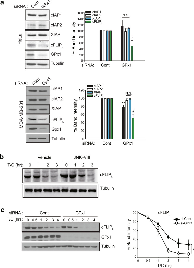
Fig. 4. GPx1 absence selectively reduces the cFLIP level.
a HeLa and MDA-MB-231 cells were transfected with the indicated siRNAs for 24 h and lysed for use in immunoblotting. The immunoblot bands were quantified and normalized to the intensity of α-tubulin bands. The data in the graph are the means ± SD of percent of the band intensities representing GPx1 siRNA-transfected cells relative to those representing control siRNA-transfected cells (n = 3, *P < 0.005, **P < 0.002, and #P < 0.001 versus the corresponding control samples). Immunoblots with α-tubulin used as the loading control. NS not significant. b HeLa cells were pretreated with the JNK inhibitor VIII (JNKi-VIII, 10 μM) for 30 min and stimulated with T/C for the indicated times. The cells were lysed for use in immunoblotting. A representative blot is shown (n = 3). c siRNA-transfected HeLa cells were stimulated with T/C and lysed at the indicated times for use in immunoblotting. The immunoblot bands for cFLIPL were quantified and normalized to the intensity of α-tubulin bands. The data in the graph are the means ± SD of percent of cFLIPL intensity in T/C-stimulated cells relative to that in unstimulated cells (n = 3, *P < 0.0001 with repeated-measures ANOVA). An immunoblot with α-tubulin used as the loading control.

