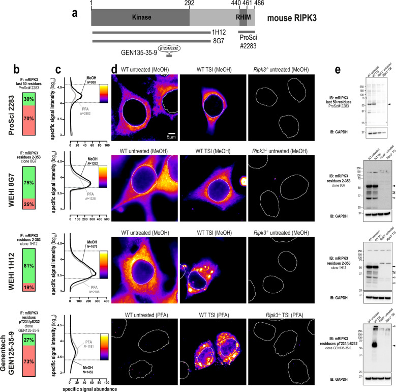
Fig. 5. Unlike human RIPK3, mouse RIPK3 is highly amenable to detection by immunofluorescence and immunoblotting.
a Mouse RIPK3 domain architecture showing the immunogens or epitopes for the tested anti-RIPK3 antibodies. b Quantitation of the percentage of gated signals for the tested RIPK3 antibodies. c Quantitation of specific signal abundance produced by the tested RIPK3 antibodies on methanol-fixed (MeOH) or paraformaldehyde-fixed (PFA) MDFs. The number of cells imaged (N) to generate each signal-to-noise curve is shown. d Micrographs of immunofluorescent signals for the tested RIPK3 antibodies on MDFs. As indicated by each pseudocolour look-up-table, only immunosignals within the respective gate in (c) were visualised. Data are representative of n = 3 (in-house clone 8G7 and Genentech clone GEN135-35-9) and n = 2 (ProSci 2283, in-house clone 1H12) independent experiments. Nuclei were detected by Hoechst 33342 staining and are demarked by white outlines in micrographs. e Immunoblot using the tested RIPK3 antibodies against lysates from wild-type and Ripk3−/− MDFs. Closed arrowheads indicate the main specific band. Open arrowheads indicate other specific bands of interest. Immunoblots were re-probed for GAPDH as loading control.

