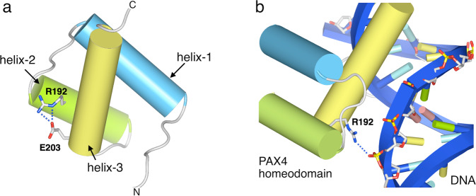
Fig. 1. Structural model of the homeodomain in human PAX4.
a The human PAX4 homeodomain is represented as cylinders, with the Arg192 and Glu203 residues being shown as sticks. The PAX4 homeodomain consists of three helices: helix-1 (light-blue), helix-2 (green), and helix-3 (yellow). Arg192 located on helix-2 is predicted to form salt-bridges with Glu203 located on helix-3. b Binding of the human PAX4 homeodomain to double-stranded DNA. DNA is shown by ribbon representation in blue. Arg192 of PAX4 (sticks) is predicted to bind with the phosphate backbone (sticks) of DNA.

