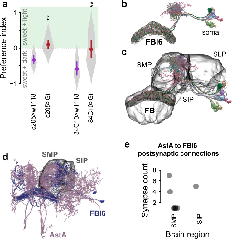
Fig. 5. FBl6 neural activity is context-dependent.

a Food preference index of flies with spatially restricted persistent optogenetic inhibition. FBl6 neurons are optogenetically inhibited in one half of the arena that contains only sweet food on both sides. This inhibition abolishes aversion for bright green light by shifting food preference away from dark half of the arena. Points on graphs depict mean ± 95% CI, with violins depicting full data distribution. b EM reconstruction of FBl6 neurons shows neural projections restricted to layer 6 of the fan-shaped body. EM reconstruction of traced FBl6 neurons from the Janelia hemibrain dataset was performed using neuPrint and NeuronBridge for neurons targeted by 84C10-GAL4 and embedded in the surface representation mesh of standardized FBl6 brain region. c EM reconstruction of FBl6 neurons targeted by 84C10-GAL4 was performed as previously and embedded in the surface representation mesh of standardized whole fan-shaped body (FB). FBl6 neural projections to superior medial protocerebrum (SMP), superior intermediate protocerebrum (SIP), and superior lateral protocerebrum (SLP) higher brain regions are shown embedded in the surface representation of these standardized higher brain regions. d AstA neurons make postsynaptic connections with FBl6 neurons in SMP and SIP brain regions. Surface representations of brain regions where AstA neurons make postsynaptic connections with FBl6 neurons are shown in gray. EM analysis of traced AstA neurons that make postsynaptic connections with FBl6 neurons was performed in neuPrint. e Quantification of number of postsynaptic connections from AstA to FBl6 neurons. Each gray dot represents the number of synaptic connections between one AstA and one FBl6 neuron, separated by brain region on the x-axis where these connections are made.
