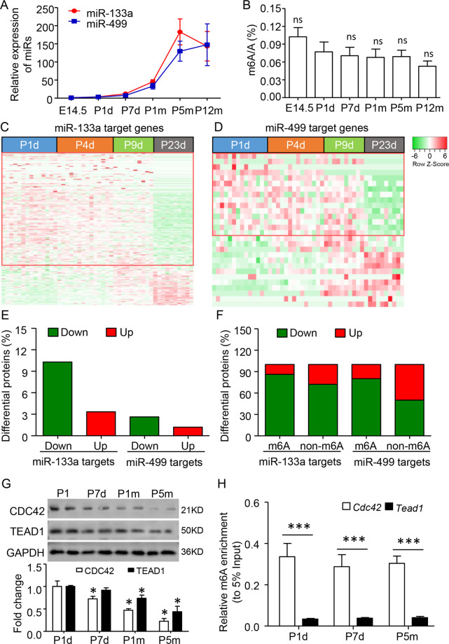
Fig. 2. m6A modification promotes miR-133a repression in heart development.
A Developmental expression profiles of miR-133a and miR-499. The expression changes were calculated by comparing with E14.5 data. B Developmental global m6A modifcation levels, as determined by ELISA analysis. ns, no significant as compared to P1d. C, D Heatmap showing the developmental changes of miR-133a and miR-499 target protein expression from the published proteomics database. The ventricular tissues were collected from the postnatal day 1 (P1) to P23 of the C57BL/6 mouse. E The percentage of the down- and upregulated proteins (Ratio of P23d to P1d) by miR-133a and miR-499. F The proportions of down- and upregulated genes targeted by two miRs with or without m6A modification. G Quantitative western blot analysis showed that the decreased CDC42 and TEAD1, two miR-133a target proteins in the developing ventricle from P1d to postnatal 5 months (P5m). Error bars, SD (n = 4–5/stage), *P < 0.05 as compared to P1d. H Enrichment of m6A modification at Cdc42 and Tead1 was analyzed by m6A-RNA immunoprecipitated-qPCR method. Data are mean ± SD (n = 4–6 per stage). *P < 0.05; ***P < 0.001 was calculated using the one-way ANOVA followed by Tukey’s test.

