Fig. 2.
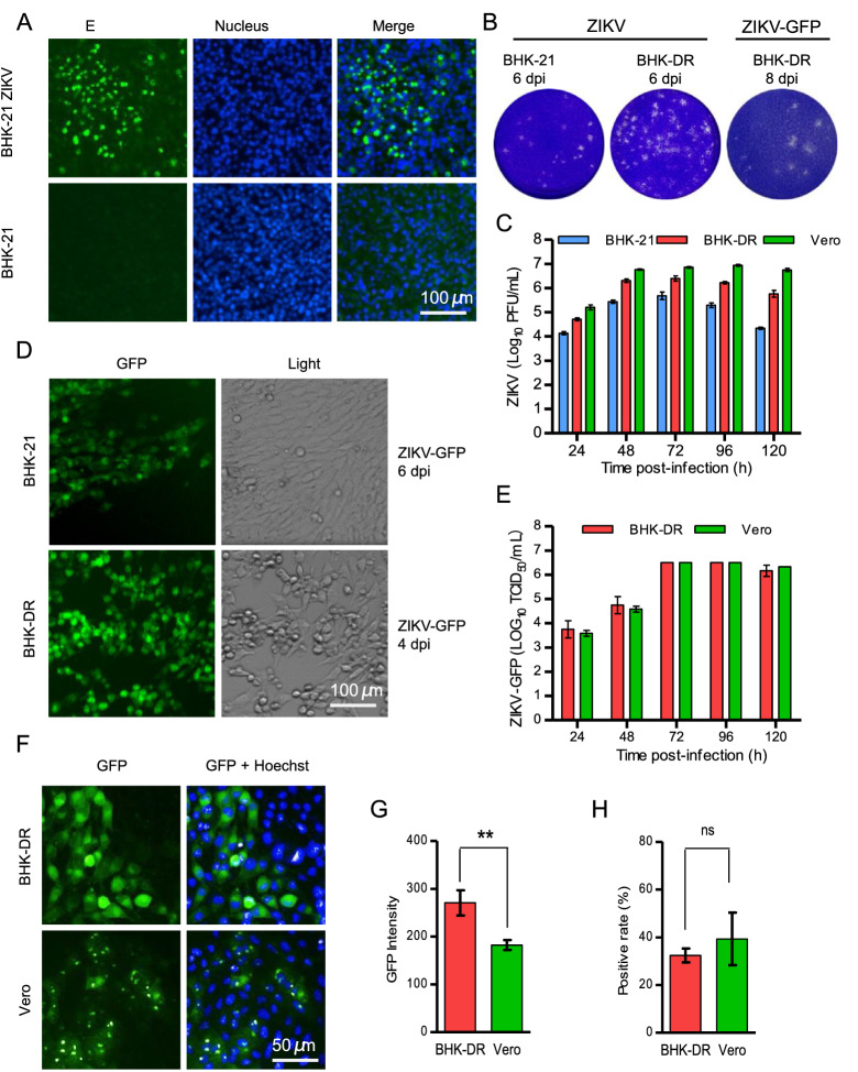
Production and characterization of recombinant ZIKV and ZIKV-GFP reporter viruses. A Immunofluorescence analysis of recombinant ZIKV. The supernatants of transfected 293T cells were inoculated into BHK-21 cells. Forty-eight hours later, infected cells were fixed and probed with pan-flavivirus E proteinspecific MAb 4G2. Nuclei were stained with DAPI. B Representative images of plaque assays of BHK-21 and/or BHK-DR cells infected with ZIKV and ZIKV-GFP viruses and fixed and stained at the indicated days postinfection. C Replication kinetics of recombinant wild-type ZIKV in BHK-21, Vero and BHK-DR cells. Cells were infected at an MOI of 0.1. Supernatants were harvested at 24 hpi to 120 hpi and analyzed using a plaque form assay in BHK-21 cells. D ZIKV-GFP reporter virus infection in BHK-21 and BHK-DR cells. Cells infected with ZIKV-GFP at the indicated time were analyzed by fluorescence microscopy for GFP expression and by light microscopy for cytopathic effects. E Replication kinetics of recombinant ZIKV-GFP in Vero and BHK-DR cells. Cells were infected at an MOI of 0.1. Supernatants were harvest at 24 hpi to 120 hpi and analyzed using a TCID assay in BHK-DR cells by observation of fluorescent foci. F GFP fluorescence in Vero and BHK-DR cells infected with ZIKV-GFP. One representative experiment out of three is shown. At 48 hpi, cells were stained with DAPI and visualized using a high-content screening microplate imaging reader (Operetta; PerkinElmer). The GFP fluorescence intensity G and GFP-positive ratio H were analyzed using Acapella high-content imaging and analysis software. The results are the mean of three independent experiments performed in triplicate. Statistical significance was determined by the Student’s t-test (** P < 0.01, ns P > 0.05).

