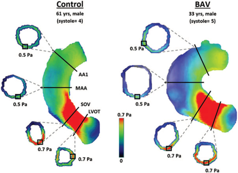
Figure 4.

Visualized examples of localized maximum WSSM values at each aortic location in a healthy control and a BAV patient. AA1, proximal to first aortic branch; BAV, bicuspid aortic valve; LVOT, left ventricular outflow tract; MAA, mid-ascending aorta; SOV, sinuses of Valsalva; WSS, wall shear stress; WSSM, magnitude wall shear stress; yrs, years old.
