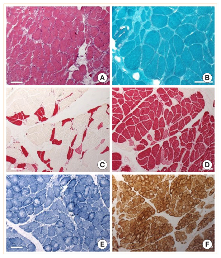
Fig. 1.
Biopsy of left musculus gastrocnemius of a patient with florid Cushing syndrome. (A) H&E stain reveals disseminated atrophic muscle fibres with mild fibrosis and increase in connective tissue. Myonucleii are somewhat more internalised and single fibres show subsarcolemmal vacuolization. (B) Trichrome Gömori staining displays no protein aggregation or ragged red fibres. Anti-myosin fast (C) and slow (D) stainings show a pronounced type-2 fibre atrophy; however, some type-1 fibres are also atrophic. Oxidative stainings (E, nicotin amide adenine dinucleotide [NADH]; F, cytochrome C oxidase/succinate dehydrogenase [COX/SDH] double staining) shows central reduction of oxidative enzymes in some fibres, pointing at an energy level alteration in those myofibres (×25).

