Fig. 1—
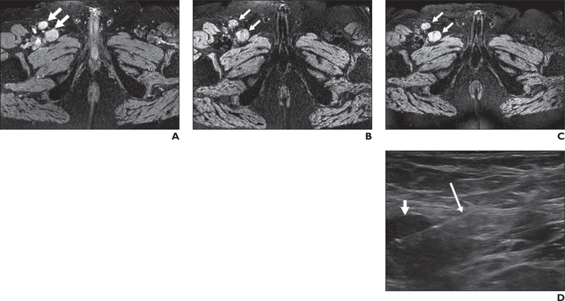
67-year-old man with prostate cancer (serum prostate-specific antigen level, 47.63 ng/mL).
A, Axial T2*-weighted MR image obtained before ferumoxytol injection shows right inguinal adenopathy (arrows).
B and C, T2*-weighted MR images obtained 24 (B) and 48 (C) hours after ferumoxytol injection show lack of ferumoxytol uptake within right inguinal adenopathy (arrows).
D, Sonogram obtained during sonography-guided biopsy shows right inguinal adenopathy (short arrow). Long arrow shows biopsy needle. Histopathology results revealed poorly differentiated prostate adenocarcinoma.

