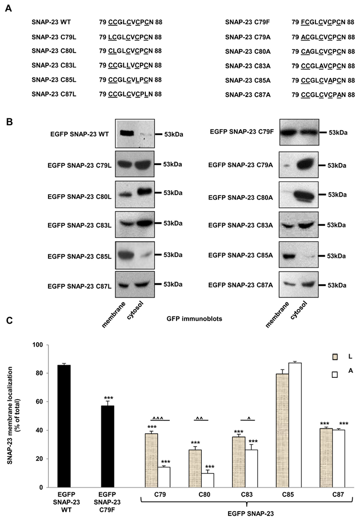
Fig. 4.

SNAP-23 single cysteine mutants show differential membrane association in RBL mast cells. A. Amino acid sequence of the cysteine-rich domain of SNAP-23 and of the corresponding region of SNAP-23 single cysteine mutants. B. RBL cells were transfected with EGFP-tagged SNAP-23 WT and various SNAP-23 single cysteine mutants. Twenty-four hours post-transfection the cells were fractionated for membrane/cytosol enrichment analysis Equivalent portions of each fraction were analyzed by immunoblotting with anti-SNAP-23 and anti-GFP antibody. Shown here are representative equivalent membrane/cytosol enrichment fraction blots out of three independent transfection- membrane/cytosol enrichment fractionation experiments for each SNAP-23 mutant. All blots for specific mutants were developed, and quantitated similarly. Each time for one PVDF membrane/blot, several films were developed with different exposure times (5 s to 1 min) for best visualization of the bands. Here the representative blots are shown with the molecular weight markers. C. Band intensities from three independent experiments were quantified by densitometry and expressed as percentage of SNAP-23 membrane localization with respect to total SNAP-23 (mean ± SEM). *** indicate statistically significant decrease in membrane binding of the mutants compared with wild-type SNAP-23 (p < 0.0005). Carets indicate statistically significant differences in membrane binding of SNAP-23 cysteine to leucine (L) and cysteine to alanine (A) mutants (^ ^ ^, p < 0.0005; ^ ^, p < 0.005 and ^, p < 0.05).
