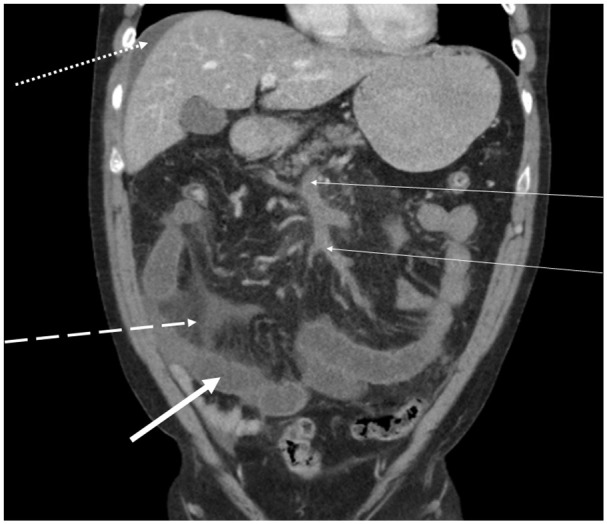
Fig. 1.

A CT with intravenous contrast enhancement in the portal phase showed MVT (two thin arrows). Note the secondary intestinal abnormalities such as dilated small bowel loops (thick arrow), mesenteric edema (dashed line), and ascites (dotted line).
