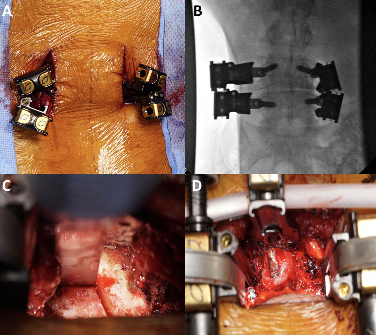
Figure 2.
Intraoperative images of minimally invasive transforaminal lumbar interbody fusion: A. Bilateral mini-open paramedian incisions are made. Pedicle screws are placed under image guidance. A screw shank/blade assembly is inserted at each level. B. Intraoperative posteroanterior fluoroscopic X-ray demonstrating the screw shank/blade assembly. C. Complete facetectomy is performed on each side using an osteotome and Kerrison rongeur. D. Central decompression is achieved by removing the ligamentum flavum and undersurface of the lamina to allow for passage of a curved freer across the midline.

