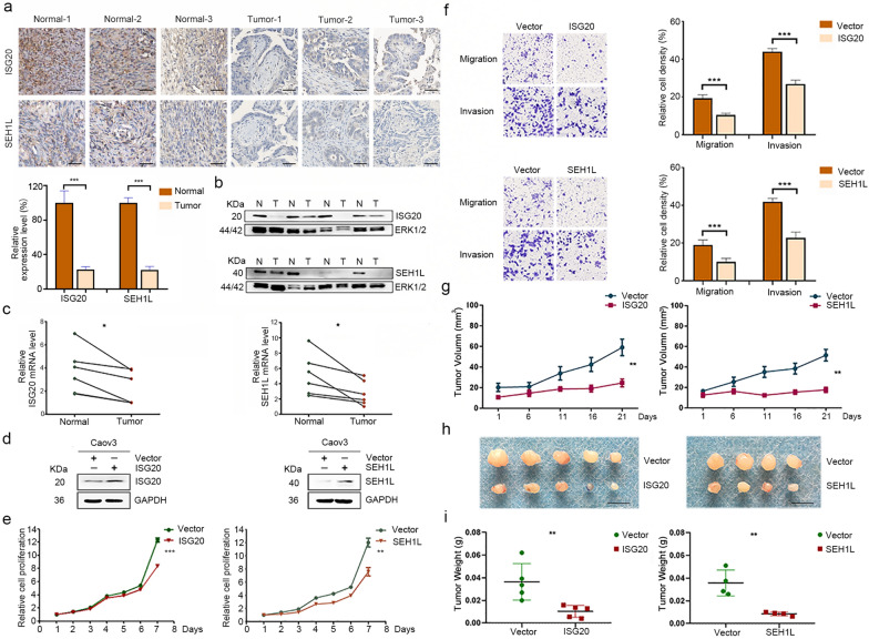
Fig. 7.
Expression and function analysis of unreported signature genes ISG20 and SEH1L in OC. a, b Immunohistochemistry (a) and western blotting (b) analysis of ISG20 and SEH1L protein levels in paired tumor tissues and adjacent normal tissues. Scale bar = 50 μm. c qRT-PCR analysis of ISG20 and SEH1L mRNA levels in paired tumor tissues and adjacent normal tissues. *P < 0.05. d Western blotting analysis of ISG20 or SEH1L expression in Caov3 cells transfected with pLVX-ISG20-IRES-Neo, pLVX-SEH1L-IRES-Neo or empty vector. e Cell proliferation was detected by the CCK-8 assay. **P < 0.01; ***P < 0.001. f Cell migration and invasion were evaluated by Transwell assays. g Tumor growth curve. h. Xenograft tumors. Scale bar = 1 cm. i The weights of xenograft tumors. **P < 0.01; ***P < 0.001

