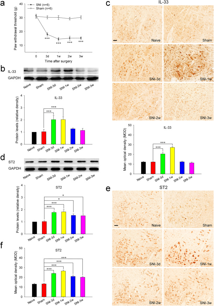
Fig. 1.
Increased expressions of IL-33 and ST2 in the RN of SNI rats. A Mononeuropathic pain induced by SNI (n = 6 per group, F = 198.886, P < 0.001). B Western blotting showed that red nucleus IL-33 was increased at 3 days, peaked at 1 week and returned to normal level at 2 weeks post-SNI (n = 6 per group, F = 9.435, P < 0.001). C Immunohistochemistry indicated that red nucleus IL-33 was upregulated at 3 days, peaked at 1 week and returned to normal level at 2 weeks post-SNI (n = 4 per group, F = 50.817, P < 0.001). D Western blotting showed that red nucleus ST2 was increased at 3 days, peaked at 1 week, and still remained at a high level at 3 weeks post-SNI (n = 6 per group, F = 7.693, P < 0.001). E, F Immunohistochemistry (n = 4 per group) indicated that red nucleus ST2 was upregulated at 3 days, peaked at 1 week, and still kept at a high level at 3 weeks post-SNI (n = 4 per group, F = 39.934, P < 0.001). *P < 0.05 and ***P < 0.001. Scale bars = 50 μm

