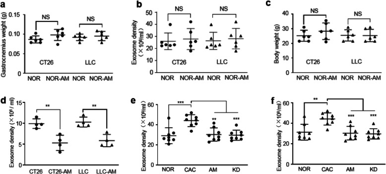
Fig. 4.
Amiloride inhibited tumor-derived exosome release both in vitro and in vivo. a Gastrocnemius weights of the NOR and NOR-AM mice in the CT26/LLC models (n = 6). b Plasma exosome densities of the NOR and NOR-AM mice in the CT26/LLC models (n = 6). c Body weights of the NOR and NOR-AM mice in the CT26/LLC models (n = 6). d Exosome densities in culture media of the CT26, CT26-AM, LLC, and LLC-AM cells (n = 4). Cells were treated by amiloride at 10 μM for 6 h. e Plasma exosome densities of the NOR, CAC, AM, and KD mice in the CT26 model (n = 8). f Plasma exosome densities of the NOR, CAC, AM, and KD mice in the LLC model (n = 8). Statistical significances: p > 0.05, NS; p < 0.05, *; p < 0.01, **; p < 0.001, ***. NOR, C57BL/6 or BALB/c normal control mice; NOR-AM, amiloride-treated NOR mice; CAC, CT26/LLC cachexia mice; AM, amiloride-treated CT26/LLC mice; KD, mice inoculated with Rab27-knockdown CT26/LLC cells; CT26-AM, amiloride-treated CT26 cells; LLC-AM, amiloride-treated LLC cells

