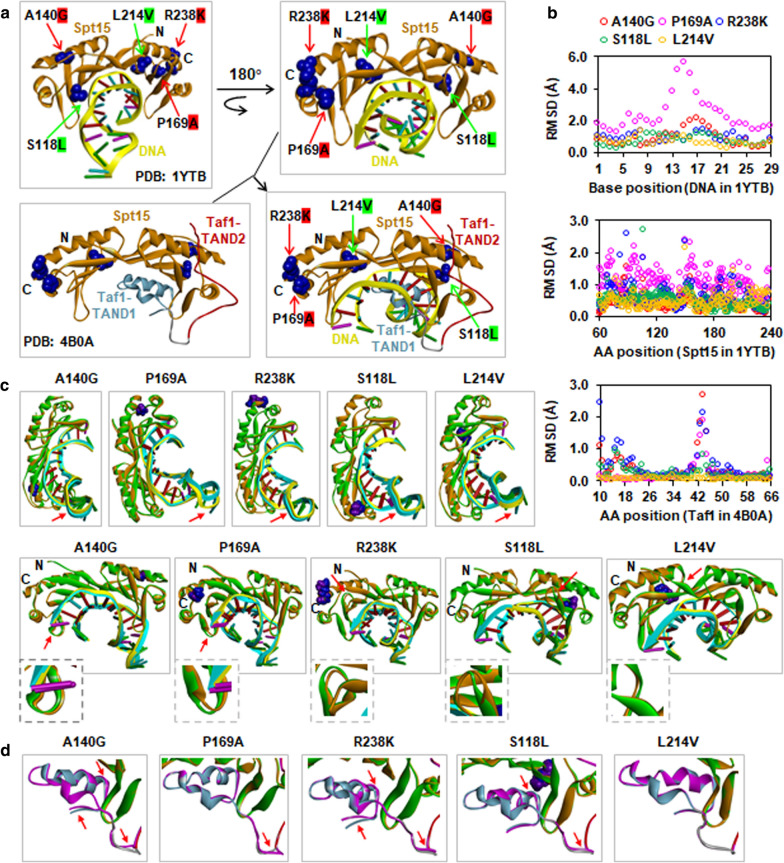
Fig. 7.
Predicted effects of key point mutations on protein conformation of Spt15 and its interactions with DNA and Taf1. a Localization of key point mutation of Spt15 on PDB structures 1YTB and 4B0A including three most stress tolerant (A140G, P169G, R238K) and two most stress-sensitive (S118L, L214V) mutations. Superposition structure of the Spt15-DNA (Chain A and C) from the structure 1YTB and Spt15-Taf1 from the structure 4B0A was constructed using super-alignment in the PyMol program. b RMSD (root mean square deviation) profiles of DNA and Spt15 in 1YTB and Taf1 in 4B0A influenced by key Spt15 mutants. c DNA and Spt15 conformation changes in 1YTB. d Taf1 conformation changes in 4B0A. Conformation changes are indicated by red arrows. The coloring of chains and residues is as follows. Wild-type and mutant Spt15 were shown in orange and green, respectively. DNA interacting with wild-type and mutant Spt15 are in yellow and cyan, respectively. In Taf1 interacting with wild-type Spt15, the TAND1 and TAND2 domains are shown in sky blue and red, respectively. Taf1 interacting with mutant Spt15 is shown in purple. Wild-type and mutant amino acids were shown in blue and purple, respectively

