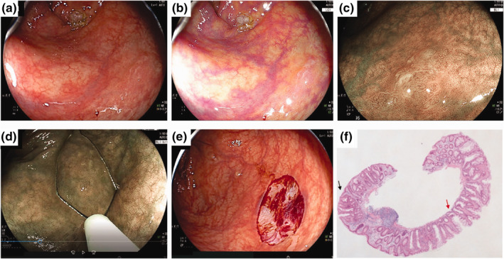
FIGURE 2.

Repeat cold snare polypectomy (CSP) for a recurrent sessile serrated lesion (SSL). (a) A non‐polypoid SSL 10 mm in size located in the ascending colon was detected by LED endoscopy (no. 3 in Table 3). (b) Linked colour imaging detected a clear lesion. (c) The lesion showed dilated crypts on magnifying endoscopy with blue laser imaging (BLI). A minor network was seen, which might have represented a small amount of dysplasia. (d) Repeat CSP was performed with a dedicated snare. (e) The lesion was resected en bloc. (f) Histopathology showed SSL with low‐grade dysplasia (black arrow). The horizontal margin of the lesion was negative, but the vertical margin was unclear (red arrow)
