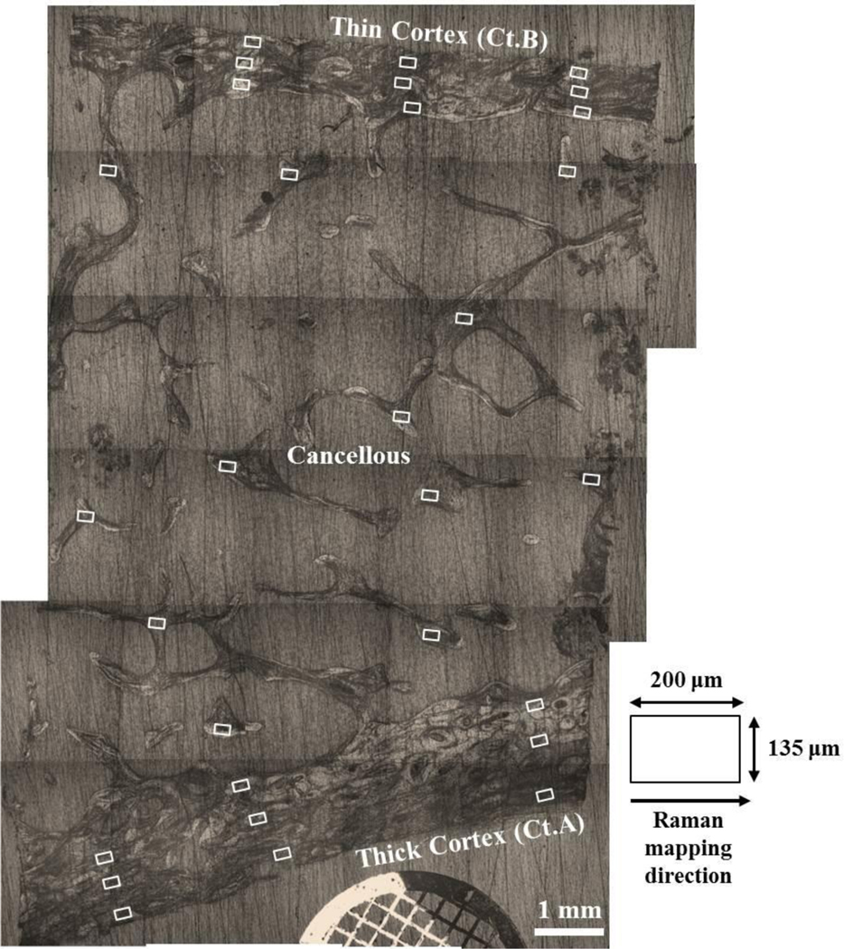
Figure 2.

Reconstructed longitudinal image of an iliac crest biopsy section showing the approximate surface locations of all 18 cortical and 12 cancellous Raman maps (white boxes). The laser line is ~135 μm in length and is aligned perpendicularly to the thick cortex (Ct.A) and the thin cortex (Ct.B) prior to Raman mapping.
