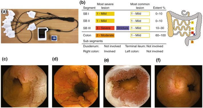
FIGURE 1.

(a) PillCam Crohn's capsule, DR3 data recorder and wireless sensors. (b) A representative graphic of a patient with active Montreal L3 B2 disease and images of small bowel (SB) lesions (c and d), SB stricture (e) and colonic lesion (f). RAPID™ Reader Software breaks down small bowel and colonic segments based on identified anatomical landmarks. The reader classifies the most severe and most common lesion (none, mild, moderate and severe), presence or absence of stricture and extent of disease (0%–10%, 10%–30%, 30%–60%, 60%–100% of segment)
