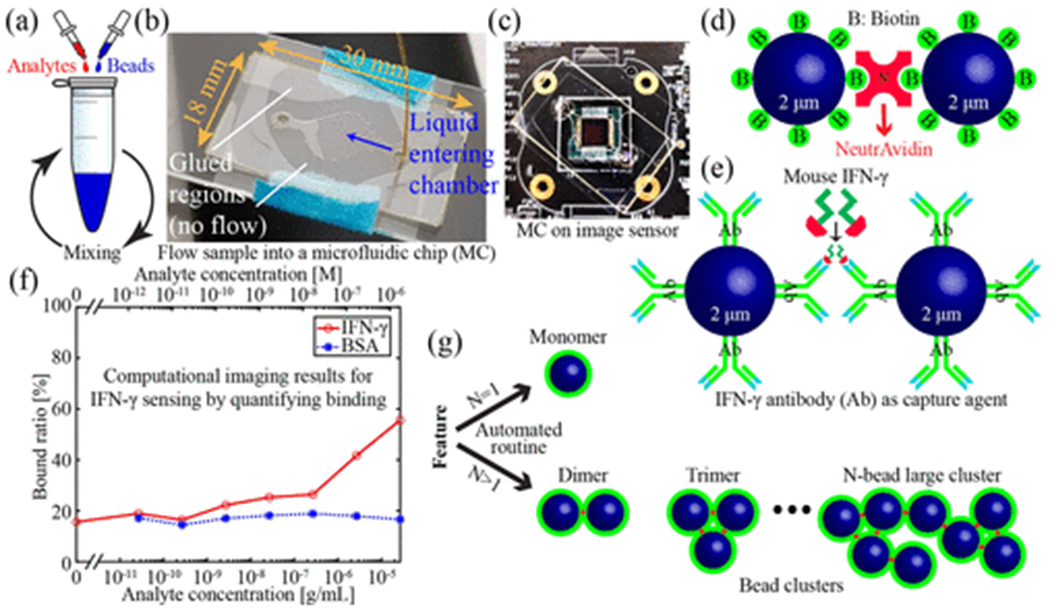
Figure 1:

Quantitative large area binding assay. (a) Incubation of analytes and functionalized beads. (b) Photograph of sample being delivered into a MC. (c) Photograph of a MC sitting on a CMOS sensor. (d) Illustration of binding between two biotin coated beads and a NA molecule. (e) Illustration of binding between two anti-mouse-IFN-γ coated beads and a mouse IFN-γ molecule. (f) Computational imaging-based sensor response for sensing mouse IFN-γ. Bovine serum albumin is used as a negative control. (g) Illustration of monomers and clusters.
