Figure 1.

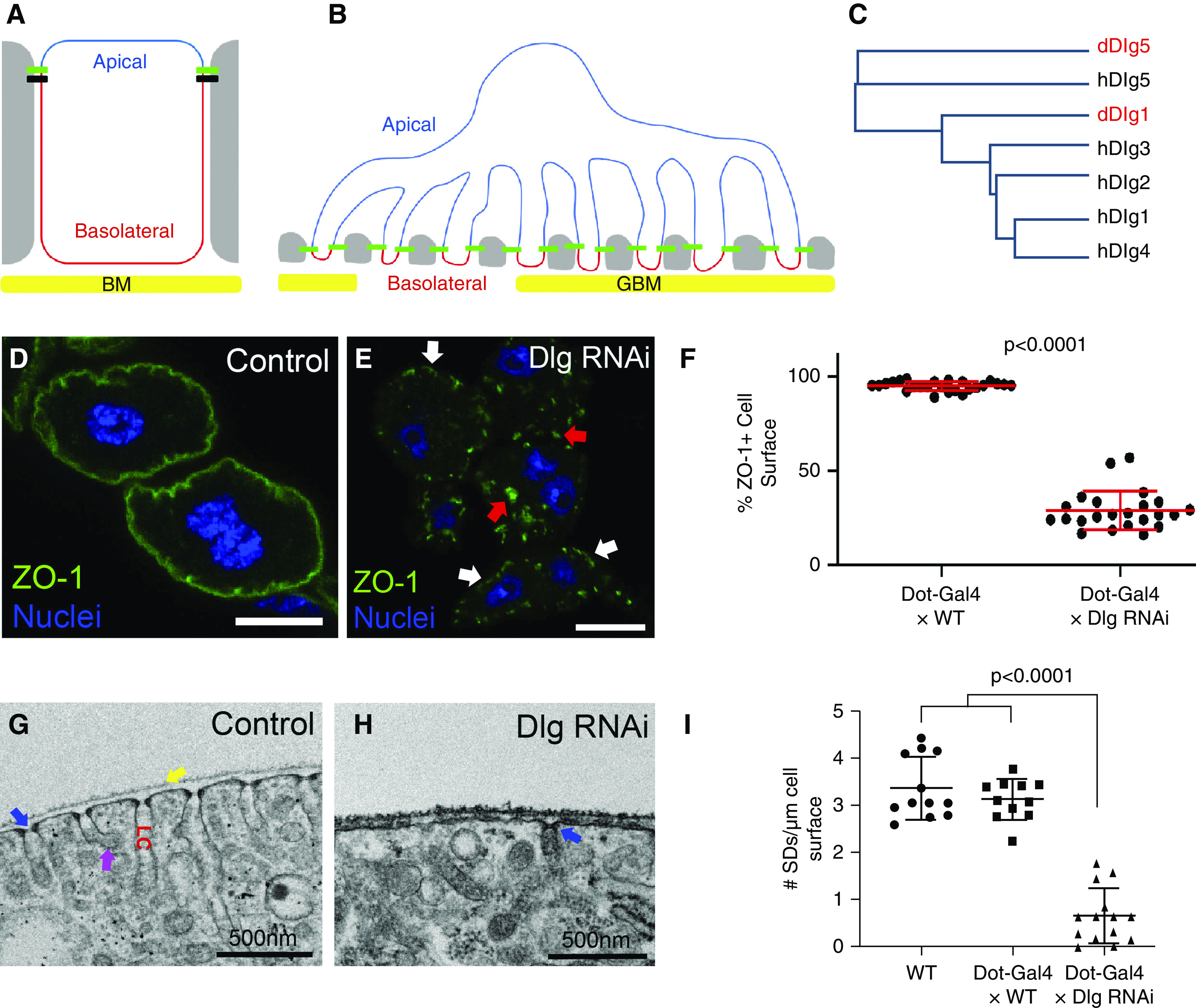
Basolateral polarity proteins promote SD formation. (A) Diagram of a classic polarized epithelial cell, such as an immature podocyte precursor, highlighting the apical domain (blue) and the basolateral domain (red), which abuts the basement membrane (BM; yellow). Tight junctions (green) and adherens junctions (black) connect to neighboring cells (gray). The apical domain in epithelia typically contains polarity proteins such as Crb, aPKC, Par3, and Par6; whereas the basolateral polarity module (Dlg, Scrib, and Lgl) localizes to the basolateral domain. (B) The complex architecture of a mature podocyte retains some aspects of apicobasal polarity, although the apical domain (blue) is now greatly expanded and the basolateral domain (red) is restricted to the region below the SDs (green), adjacent to the GBM (yellow). SDs form the intercellular junctions connecting foot processes of the featured podocyte to foot processes of neighboring podocytes (gray). (C) Dendrogram based on protein sequence alignment of the five human and two Drosophila Dlg proteins; fly Dlgs indicated in red. Human Dlg1 to 4 proteins are most similar to fly Dlg1. Both human and fly Dlg5 proteins cluster separately. (D) Control nephrocyte (Dot-Gal4×WT) stained for the SD protein ZO-1 (green) shows uniform localization along the entire surface of the cell (95.3% of cell surface stains positive for ZO-1; n=25). Note that garland nephrocytes are binucleate (blue). (E) KD of fly Dlg (Dot-Gal4×Dlg RNAi) significantly disrupts ZO-1 localization along the surface of the nephrocyte (only 29.1% of cell surface is ZO-1+; n=23; P<0.001 compared with control); graphed in (F). Small regions of cell surface retain ZO-1 localization (white arrows), whereas numerous internal aggregations can be found in the cytoplasm (red arrows). (G) TEM showing a cross-sectional view of a small surface region from a control nephrocyte. SDs are regularly spaced across the entire surface of the cell; one SD is highlighted by the blue arrow. Nephrocytes are covered with a basement membrane (yellow arrow). Internal to the SDs are invaginations of the PM known as labyrinthine channels (LC). The membrane along these channels is highly endocytic, with many clathrin-coated vesicles budding off (magenta arrow). (H) Consistent with our ZO-1+ surface localization analysis, TEM images of Dlg-KD cells show significant reduction in the number of SDs (blue arrow); quantified in (I). Error bars represent mean and standard deviation. Scale bars, 10 μm.
