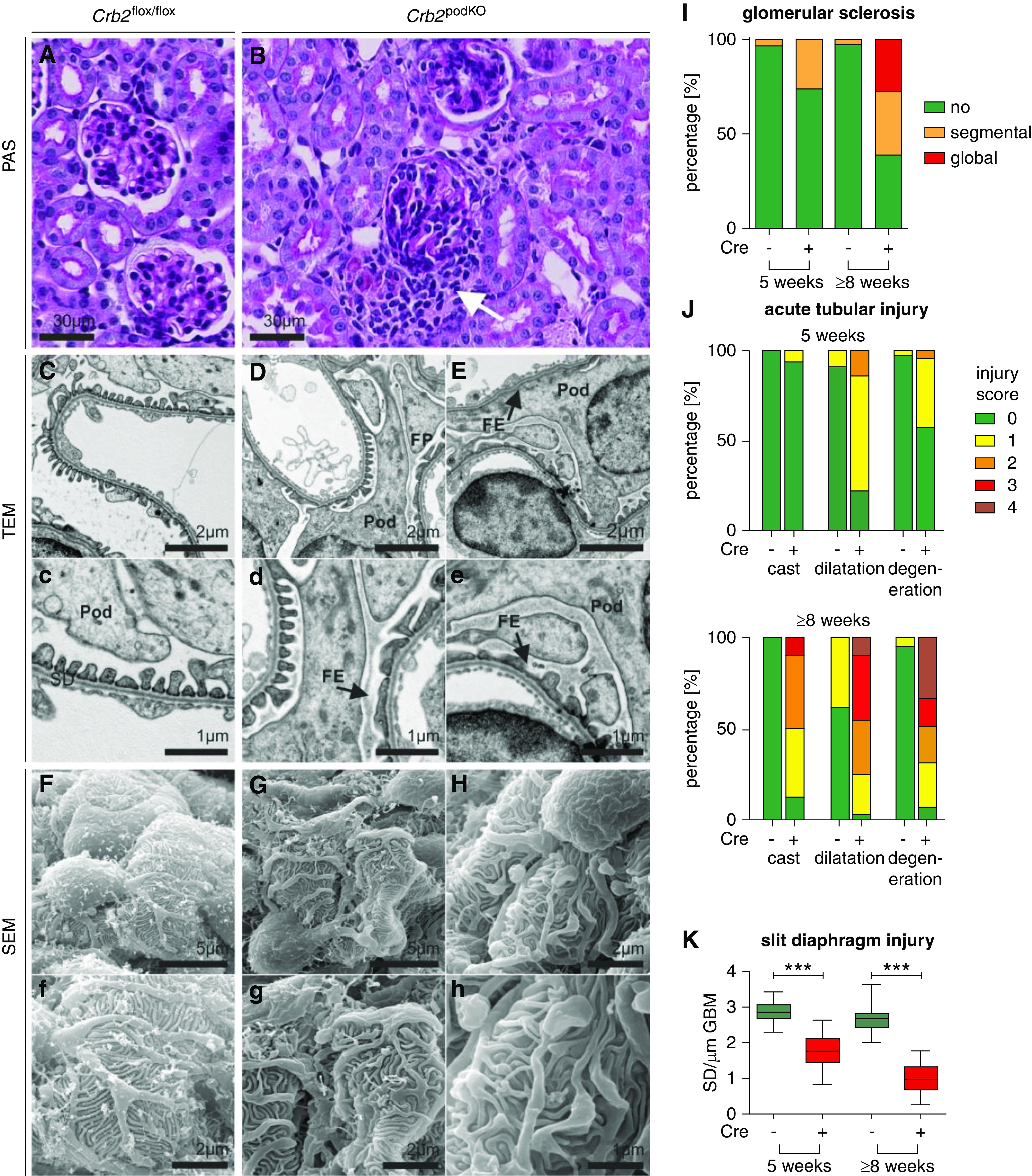
Figure 2.

Crb2 loss results in disordered and effaced foot processes (FPs). (A and B) Periodic acid–Schiff (PAS) staining. Kidney tissue of 5-week-old Crb2 podKO infiltration of cells (white arrow). (C and D) TEM analyses. Ultrastructural analyses revealed segmental foot process effacement (FE; black arrows) in (D, d, E, and E) Crb2 podKO in comparison with (C and c) Crb2 flox/flox littermate control mice. (F–H) Scanning electron microscopy analyses. (F and f) In contrast to Crb2 flox/flox, (G, g, H, and h) podocyte FPs of Crb2 podKO mice were disordered and variable in size. Pod, podocyte cell body. (I–K) Quantification of the histopathologic changes in 5- and ≥8-week-old animals revealed increased kidney injury in Crb2 podKO mice (n=3–5) (Supplemental Tables 5 and 6). (I) Percentage of glomeruli (n≥3 animals, ≥70 glomeruli) with segmental, global, or no glomerular sclerosis. (J) Percentage of the corticomedullary area (n≥3 animals) with a given acute tubular injury score with respect to tubular cast formation, tubular dilation, and tubular degeneration. (K) Quantification of slit diaphragm number in TEM images of 5- and 8-week-old mice showed strong reduction of slit diaphragm number in Crb2 podKO as compared with Crb2flox/flox mice (n=3–4). Unpaired two-tailed t test. SD, slit diaphragm. ***P<0.001.
