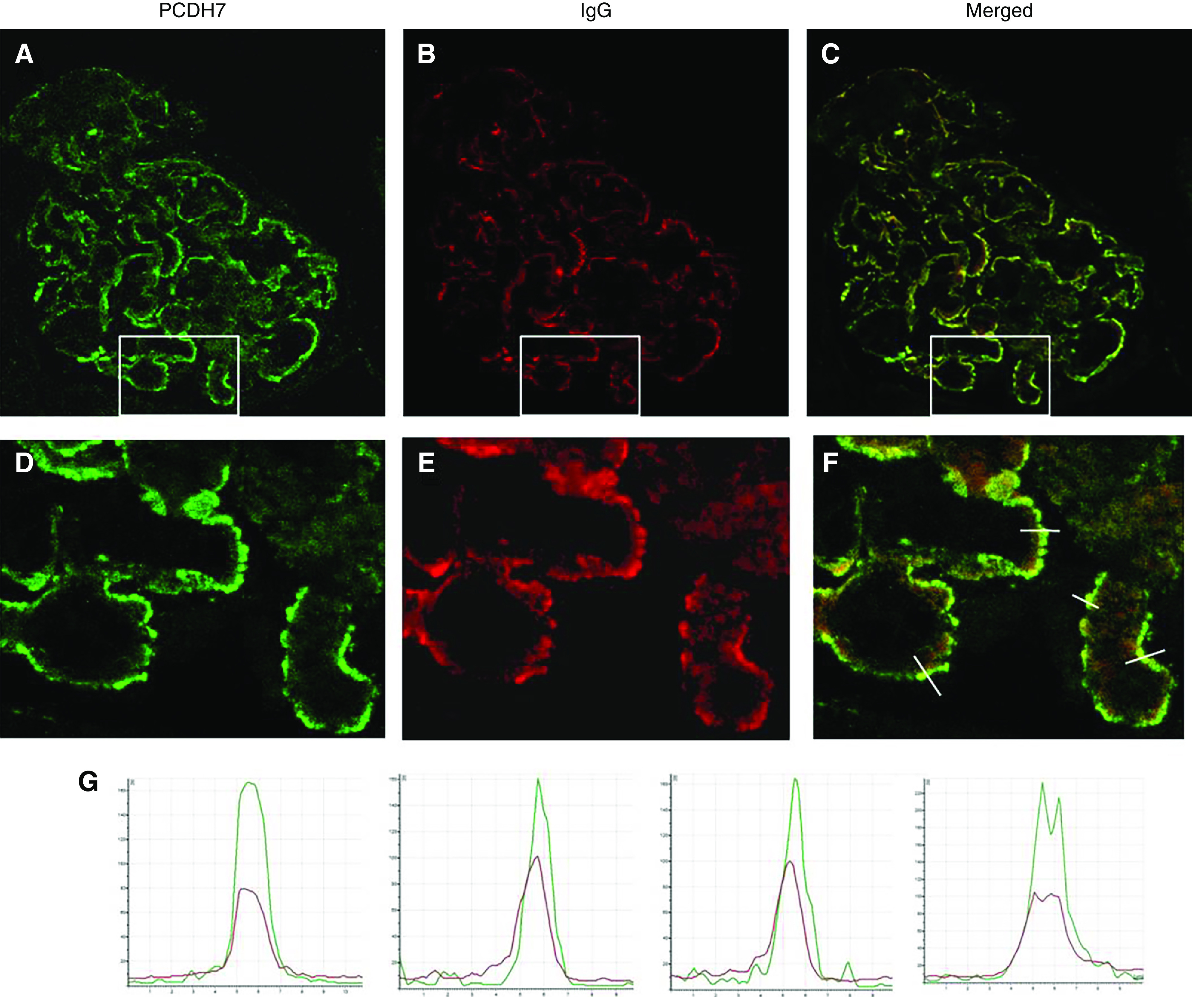
Figure 4.

Confocal IF analysis: detection of PCDH7 and IgG in glomerular immune deposits in PCDH7-associated MN. Glomeruli double labeled with (A) anti-PCDH7 (green) and (B) anti-human IgG (red). (C) The merged image. (D–F) These images are enlarged images of the boxed areas in A–C, respectively. (G) The graphs show quantitative analyses of the fluorescence recorded across sections of a representative capillary loop (indicated by lines in (F)). Note the superimposition of the two signals, which indicates that subepithelial immune deposits contain PCDH7 (green) and IgG (red).
