Figure 5.

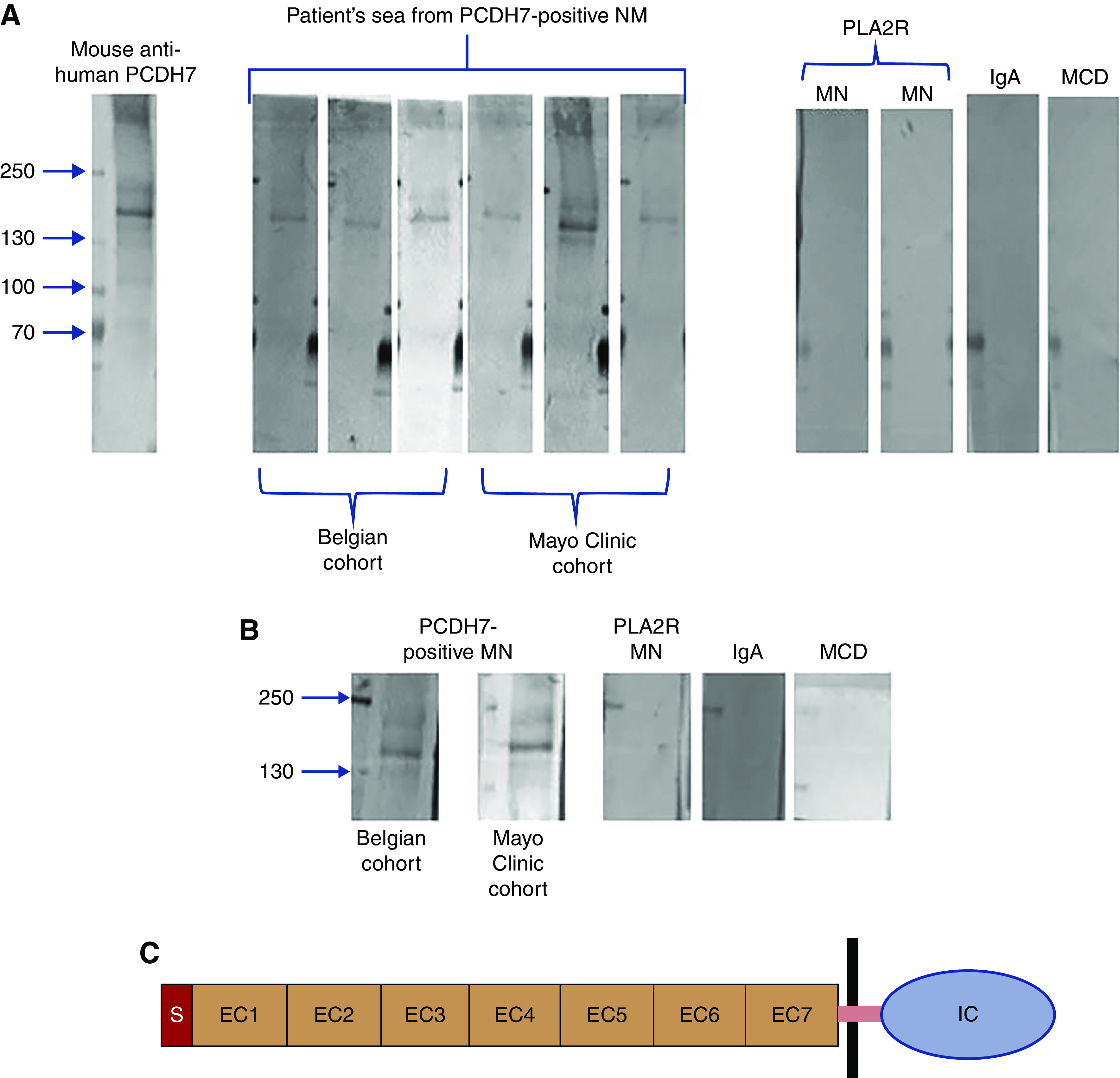
Detection of anti-PCDH7 protein antibodies by western blot analysis. (A) Under nonreducing conditions, PCDH7 was detected using mouse anti-human PCDH7 antibody as a band at 140 kD. The same band was recognized by sera from patients with PCDH7-associated MN but not by sera from patients with other kidney diseases. Each lane shows an individual patient from the discovery cohort (Mayo Clinic) or from the Belgian validation cohort. The three Mayo Clinic cohort patients are patients 1, 9, and 10, and the three Belgian cohort patients are patients 11–13. (B) Reactivity of eluted IgG from biopsy specimens. Each lane represents the eluates from three pooled biopsies from the Belgian cohort and the Mayo Clinic cohort. The three lanes on the right represent eluates from three pooled biopsies from patients with PLA2R MN, IgA nephropathy, or minimal change disease (MCD). Only IgG eluted from the PCDH7-associated MN samples reacted with the recombinant PCDH7 protein at the expected mol wt. (C) Schematic representation of PCDH7. The cadherin family is characterized by repeating motifs of extracellular cadherin (EC) domain. PCDH7 has a signal (S) peptide, seven EC domains, a single pass transmembrane domain (purple bar), and an intracellular cytoplasmic (IC) domain.
