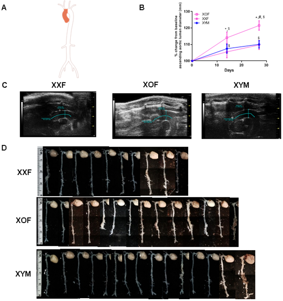
Figure 1.

XOF C57BL/6 mice have dilated ascending aortas when infused with AngII. A, Aorta illustration with shaded area depicting ascending aorta. B, Percent change from baseline of ascending aortic lumen diameters of XXF, XOF or XYM mice during AngII infusions. Data are mean ± SEM from FXX=14, FXO=14, and MXY=15. C, Representative ultrasound images of ascending aorta, blue lines delineate areas of measurement. D, Representative aortas from mice of each group, the last aortas on the right within XOF and XYM groups were classified as ruptures. *, P<0.05 FXO compared to XXF. #, P<0.05 XOF compared to XYM. $, p<0.05 compared to day 0 within genotype.
