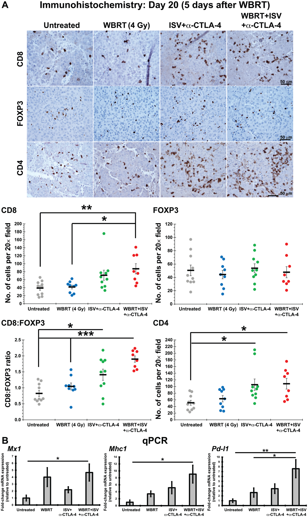
FIG. 2.

Panel A: Immunohistochemistry for T-cell markers 5 days after WBRT [4 Gy × 1, administered on day 20 of in situ vaccine (ISV) + anti-CTLA-4 (a-CTLA-4) regimen] compared to those of untreated and single-treatment controls (brown = positive immunolabeling), and quantified [***P < 0.001, **P < 0.01, *P < 0.05, mean ± SE with marker representing each individual mouse (i.e., average of 3 high-powered fields), ANOVA with post hoc Bonferroni, n ≥ 9 in at least 2 independent animal experiments]. Panel B: qRT-PCR analysis for expression of tumor immune-susceptibility genes Mx1, Mhc1 and Pd-l1 in melanoma brain tumors of standard mice at 5 days after WBRT + ISV + anti-CTLA-4, compared to those of single-treatment and untreated controls (**P < 0.01, *P < 0.05, shown as fold-change increase from untreated controls, mean ± SE, ANOVA with post hoc Bonferroni, n ≥ 8 in at least two independent animal experiments).
