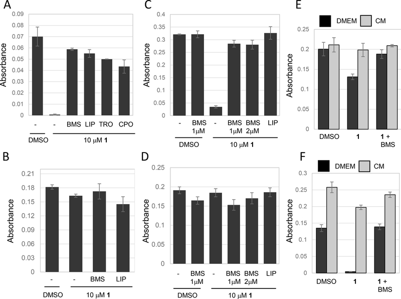
Figure 3.
Serum starvation induces resistance to ferroptosis. Cells were treated as indicated and viability determined using methylene blue 24 hours after drug treatment in all experiments. (A) Removing serum at the time of compound 1 treatment does not block ferroptosis. NCI-H522 cells were plated in DMEM+10% FBS and the next day medium was changed to DMEM without serum. Cells were immediately treated with the compounds indicated. compound 1 was used at 10 μM, BMS536924 at 1 μM, liproxstatin-1 (LIP) at 0.25 μM, trolox (TRO) at 100 μM and ciclopirox (CPO) at 5μM. (B) Serum starvation for 24 hours blocks ferroptosis. NCI- H522 cells were incubated in DMEM without FBS for 24 hours and then treated with compound 1. Viability was determined 24 hours later. (C and D) Serum starvation also blocks ferroptosis in HT1080. Cells were switched to DMEM without serum either at the time of compound 1 treatment (C), or 24 hours before compound 1 treatment (D) and viability determined. (E and F) Conditioned media enhances ferroptosis. Conditioned medium (CM) was prepared by incubating NCI-H522 cells for either one or two days in the presence of DMEM (no serum). Next, separated plates of NCI-H522 cells were starved for 24 hours in DMEM without serum. Medium was then replaced with 1-day CM (E) or 2-day CM (F). compound 1 as then added and viability determined 24 hours later. As before, starvation reduces sensitivity to ferroptosis, however ferroptosis returns in cells exposed to 2-day CM.

