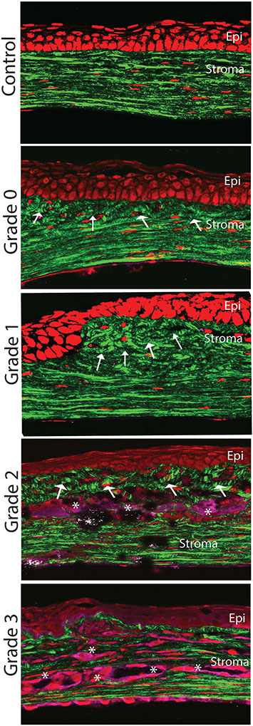
Fig. 8.
Changes in stromal organization in scars of different severity. Normal well-organized matrix and keratocytes (control normal stroma). More prominent matrix disorganization mostly localized to the anterior stroma noted in mild and moderate scars (Grades 0 and 1). Progressive worsening in matrix organization, cellular infiltration and neovascularization in more dense scars (Grades 2 and 3). Arrows show lamellae organization and asterisks presumed neovascularization.

