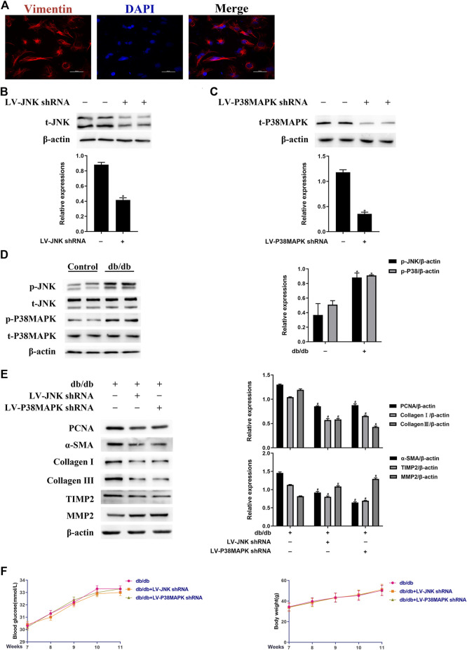
FIGURE 2.
JNK and p38 MAPK mediate the diabetes-induced increase in fibrogenic protein expression in vivo (A) Left ventricular fibroblasts isolated from mice were confirmed by vimentin staining. Data was obtained from three independent experiments (B and C) Isolated left ventricular fibroblasts from wild-type mice that were or were not injected with lentivirus via a tail vein were evaluated for JNK and p38 MAPK protein expression after 4 weeks (D) Left ventricular fibroblasts were isolated from mice and JNK and p38 MAPK phosphorylation and total JNK and p38 MAPK expression were determined by western blotting (E) Isolated left ventricular fibroblasts from diabetic mice that had or had not been injected with lentivirus via a tail vein were evaluated for PCNA, α-SMA, TIMP2, collagen I, collagen III, and MMP2 expression after 4 weeks (F) Between 7 and 11 weeks following the tail vein injection of lentivirus, the blood glucose concentration and body mass of the mice were measured. Data in (B–F) are presented as mean ± SEM (n = 6). p-values in (B–D) were calculated using paired Student’s t-test. p-values in (E-F) were calculated using one-way ANOVA with Tukey multiple comparison test. *p < 0.05, compared to the Control group; # p < 0.05, compared to the db/db mice group.

