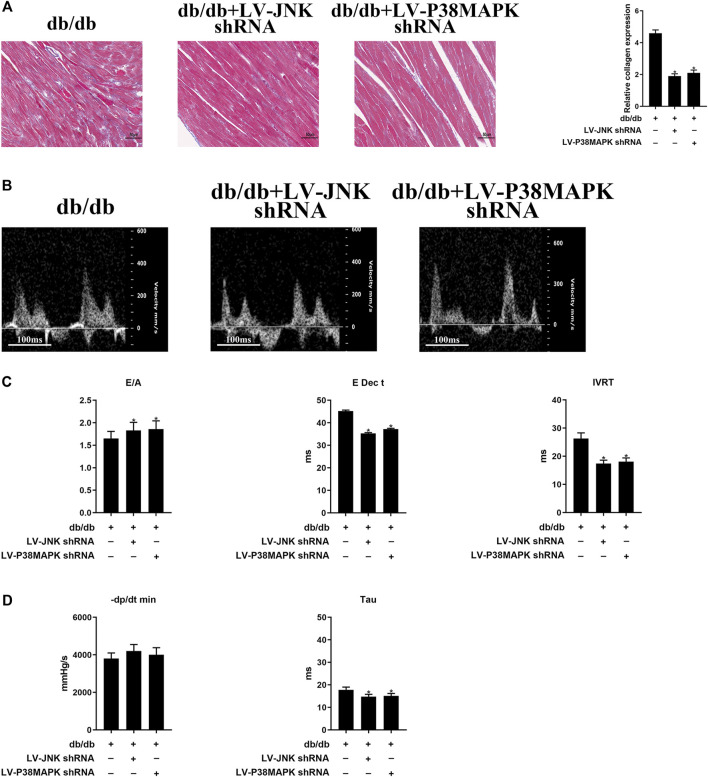
FIGURE 3.
Myocardial fibrosis and cardiac diastolic dysfunction (DD) are ameliorated by the knockdown of JNK or p38 MAPK (A) Massonʼs trichrome staining of heart tissue (original magnification ×400). The columns show the differences in collagen accumulation. Scale bars, 50 μm (B) Echo-Doppler traces for transmitral flow (C) Transmitral pulsed-wave Doppler measurements of E/A, E Dec t, and IVRT (D) Hemodynamic parameters: −dP/dt min and the time constant, Tau. The columns show the differences in left ventricular DD and hemodynamic parameters. Data are presented as mean ± SEM (n = 6). p-values were calculated using one-way ANOVA with Tukey multiple comparison test. *p < 0.05, compared to the db/db mice groups.

