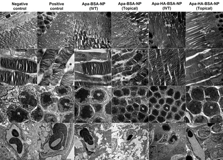
Figure 4.
Electron micrographs of retinal sections from negative control show normal organized photoreceptor cell layer (PRL), the photoreceptor cells are with regular horizontal lamellar discs (D). Outer nuclear cell layer (ONL) shows minimal intercellular spaces between nuclei (N) of photoreceptor cells. Normal blood vessel (BV) with normal thickness of the basement membrane is noticed. Electron micrographs from positive control show disorganized photoreceptor cell layer (PRL) with increased interdisc spaces (*), photoreceptor cells show distorted lamellar discs (D) and vacuolated outer segments (OS). ONL shows wide spaces between photoreceptor cells’ nuclei (N). Notice BV with thickened basement membrane. Electron micrographs from Apa-BSA-NPs (IVT) group show PRL with few distorted lamellar discs and vacuolated OS and increased interdisc spaces (*), these findings are more prominent in Apa-BSA-NPs (Topical) group. Electron micrographs from Apa-HA-BSA-NPs (IVT) and Apa-HA-BSA-NPs (Topical) groups show almost normal retinal ultra-structure. All treated groups showed normal BV basement membrane thickness.

