Figure 1.
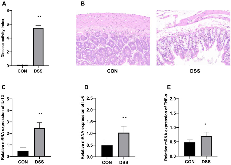
Assessment of disease activity index (A). Histopathological analysis of the colon of the control and DSS groups colitis (B), colon tissues were stained with H&E (200×magnification). mRNA expression of the proinflammatory cytokines IL-1β (C), IL-6 (D), TNF-α (E). Data are means±SD (n=7), *p<0.05, **p<0.01 compared to the control group.

