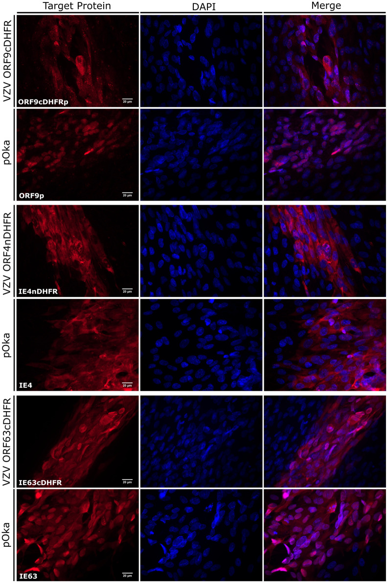
Fig 6. Localization of degron fused viral proteins compared to wild-type VZV proteins in infected cells.
(A) Images show the edges or small regions of plaques formed by wild-type VZV (pOka), VZV ORF9cDHFR (top), VZV ORF4nDHFR (middle), or VZV ORF63cDHFR (bottom) at 2-dpi. Protein cellular distribution in fixed cells was imaged to represent the distribution seen in the cultures after staining with antibodies to ORF9p, IE4, or IE63. The ‘target protein’ column indicates the specific protein probed, as noted in the lower left corner of the column. The center column shows DAPI stained nuclei, and the rightmost column shows a merged panel of target protein immunofluorescence and DAPI staining. Magnification: 60X (N.A. 1.25) oil. Single images are representations of a minimum of 15 images analyzed for each virus.

